
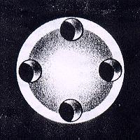
The perforated eye sphere is made of medical grade silicone elastomer. It is molded in one piece. The perforated eye sphere is available in four sizes 14mm, 16mm, 18mm, & 20mm diameter. The information given in this brochure is not exhaustive. It is meant for broad guidance only. The surgeon is advised to use the method which his own practice and discretion dictate to be best for the patient. Introduction: In patient, in whom the enucleating had been done, there is no mobile base to fix prosthesis. The purpose of perforated silicone eye sphere is to maintain contour of eye ball, maintain the eye movements, provide a base for prostheses, maintain shape of eye socket and to prevent bony deformity. Lastly, but not the least to give full self confidence to the patient. It gives excellent cosmetic results. The artificial eye prostheses, to be wore later on, will look natural due to good eye contour and almost full eye movements. How supplied: The perforated eye sphere is supplied in peel open packs. Ready to use, sterilized by gamma-rays. Each packet contains one eye sphere. The perforated eye sphere is for single use only. Indication: Perforated eye sphere is indicated in all cases where enucleating is done and good cosmetic results are desirable. Contraindications: Like all implants, it is contraindicated in cases where active infection or inflammation is present. Preferably it should be inserted after six months to one year of subsidence of inflammation. Operative procedure: Enucleating of eye ball is done on standard lines. Before division of rectus muscles retention sutures are applied as follows. A double armed suture of 1.5 metric (5/0) chromic collagen is passed through the muscle 3mm behind its insertion an transversely to the long axis of the fibers as a "whip" stitch through one edge, a mattress in the center of the muscle and a "whip" stitch at the other edge. This suture is held in pressure forceps and lifted so that the muscle is raised from the sclera to allow passage of one blade of the strabismus scissors beneath the muscle. The muscle is then divided 1mm behind its insertion. Same procedure is performed with all muscles. After removal of the eye ball and complete haemostasis a proper size of perforated eye sphere is selected. It should be smaller than the original eye ball. The pack is removed from Tenon's capsule and cavity sprayed with an antibiotic. The eye sphere is held by clean gloved hand. Preferably wash the gloved hand with sterile saline or water before touching the sphere. One of the retention sutures is passed through the hole in the eye sphere. The suture is inserted from the round end and so that it emerges through the depressed flat end of eye sphere. The idea is to keep the round side of eye sphere posteriorly and flat side anteriorly. The retention sutures of other three recti muscles are passed in similar fashion through the holes in perforated eye sphere. The sequence of rectus muscles is maintained i.e. 3 o'clock muscle suture is passed through 3 o'clock hole. In a similar fashion 6 o'clock muscle suture is passed through 6 o'clock hole. In this fashion all four retention sutures are passed through holes. Now the sphere is pushed into Tenon's capsule and ends of all four muscles are drawn out through the holes. The inferior rectus is first laid into the central depression of the sphere, where it is over-lapped for about 5mm by superior rectus. The suture in the inferior rectus passes through the deep surface of the superior rectus about 4mm behind its free end and is tied by surgical knot on the surface of the superior rectus muscle. The suture is the superior rectus muscle transfixes the edges of the inferior rectus in the form of a "whip" stitch and is then carried transversely across the united muscles to be tied by a surgical knot. A similar procedure is adopted with the other two muscles. To make it more secure. Adjacent muscles are also stitched together. The free end of the superior oblique is stretched to the medial edge of superior rectus and stitched. The free end of the inferior oblique muscle is sutured to the lower margin of the lateral rectus at the equator of sphere. The closure is performed on standard lines. Adequate pressure bandage is applied. Post operative management: The patient may be mobilized early. On the first postoperative day the conjunctiva sac is irrigated. The firm pressure dressing is maintained for 2 days, when the socket is dressed. This may be re-applied with daily dressing until the fifth day. An acrylic shell may be placed in the conjunctiva sac and a convex black eye shade, lined with sheet of lint, the smooth side opposed to the socket, is applied. The acrylic shell, approximately the shape and size of prostheses to be fitted later, helps to reduce the edema of the conjunctiva and to maintain the appropriate size and shape of the socket. A prosthesis is fitted in the third or fourth week of operation. Pain may be severe for two three days and chemosis may take up to three weeks to subside. Complications: The main complications of use of an eye sphere are infection and expulsion. To prevent infection, use of implant should be avoided with active inflammation. Suitable antibiotic cover should be given during and after implantation. Returned goods policy: Surgiwear will accept this product for replacement or credit. Provided it is returned in unopened and unsoiled packages, unless returned due to a complaint of product or mislabelling. Products will not be accepted for replacement or credit, if they have been in possession of customer for more than 90 days. Determination of product defect and mislabelling will be made by Surgiwear and will be final. Warranty: Surgiwear warrants that device has been manufactured with best quality raw material and reasonable care has been taken in manufacturing of this device. Surgiwear will not be liable for any incidental or consequential loss, damage or expense directly and indirectly arising from use of this device. The liability of Surgiwear is limited to the replacement of the product should. Surgiwear's investigation show that the product was defective at the time of its' shipment. No person has any authority to bind Surgiwear to any representation of warranty concerning this device. Autoclaving: Eye sphere is supplied sterile. If required, it can be re sterilized using following procedure: In a clean environment and with gloved hands (wash gloves with diolilled water before touching Eyesphere remove implant from its package. It should be placed directly in a stainless steel container without any covering. Autoclave for 30 minutes at 121 Celsius and 1kg/cm pressure. Any other standard autoclave cycle can also be used. Do not re-sterilize if the implant has come in contact with body fluids.
Main Products
silicone implantable devices, disposable drapes & dressings calcium hydroxyapatite (g-bone), (G-eye), fallopian tubal ring